Focus on EP | Fluoroless Ablations For Cardiac Arrhythmias

It is well known that the use of fluoroscopy during catheter ablation procedures increases the cumulative lifetime radiation exposure for both patients and operators, potentially leading to a higher risk of cancer and radiation-related injuries.1-3
Fortunately, the electrophysiology (EP) community has started to work towards minimizing the exposure to radiation during ablation procedures.
Great technological advances in EP in the last decade allow us to move toward zero or near-zero fluoroscopy ablation procedures. The use of 3D electroanatomical mapping (EAM) systems, contact-force sensing technology in combination with intracardiac echocardiography (ICE), have enabled us to visualize intracardiac catheters with a great degree of accuracy and safety.
Ablation of cardiac arrhythmias without fluoroscopy has been described and shown to be safe in the ablation of supraventricular tachycardia, typical atrial flutter and atrial fibrillation (AFib).4-7
This idea is not new. Nearly a decade ago Ferguson et al.,5 demonstrated that fluoroless ablation for AFib was safe and feasible in 21 cases; however, they required fluoroscopic rescue for two cases to aid with transseptal puncture (with two to 16 minutes of fluoroscopy for these procedures).
Advantages of Fluoroless Ablation Procedures
- No exposure to radiation for the patient, the operator and staff in general.
- No need to wear lead, reducing the risk of occupational injuries among interventional electrophysiologists.9
- Direct visualization of the intracardiac structures with ICE imaging and lesion creation.
- Similar safety and efficacy.
Potential Disadvantages of Fluoroless Ablation Procedures
- Requires the use of ICE imaging, which could be more expensive and require operator experience. Some institutions have described the use of transesophageal echocardiogram to guide transseptal puncture; however this requires a dedicated operator for image acquisition and interpretation. In addition, this is more invasive with an increased risk of complications and may not be possible in some patients.
- Modest learning curve.
It's important to highlight that rotational ICE and not phased-array ICE was used in their study, which differs from the standard phased-array ICE used in most EP procedures nowadays.
There are reports on using non-fluoroscopy ablations in outflow-tract ventricular arrhythmia (VA) ablation as well as for VA in structural heart disease from ischemic and nonischemic diseases which showed similar safety and efficacy with fluoroscopic-guided procedures, with a modest learning curve and no increase in procedural times.
Recently, a report on 1,000 consecutive patients in Europe who underwent near-zero fluoroscopic AFib ablation was published, showing that AFib ablations could be performed with very low fluoroscopy times (<six minutes) without compromising safety or efficacy.8
Our Experience
During my fellowship training at my institution, we have implemented an approach of zero or near-zero fluoroscopy for nearly all ablation procedures, under the guidance and orientation of John D. Ferguson, CHB, MB.5
This approach includes the use of more advanced technology, such as improved versions of EAM mapping systems with image integration and 3D anatomic reconstruction using the phase-array ICE integration (CartoSound Module; Biosense Webster) and the use of contact-force sensing catheters.
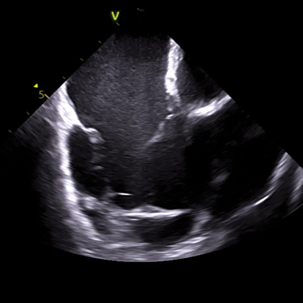
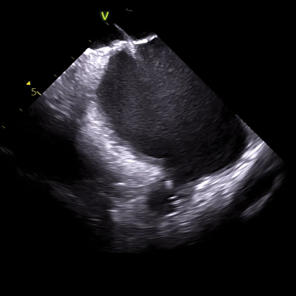
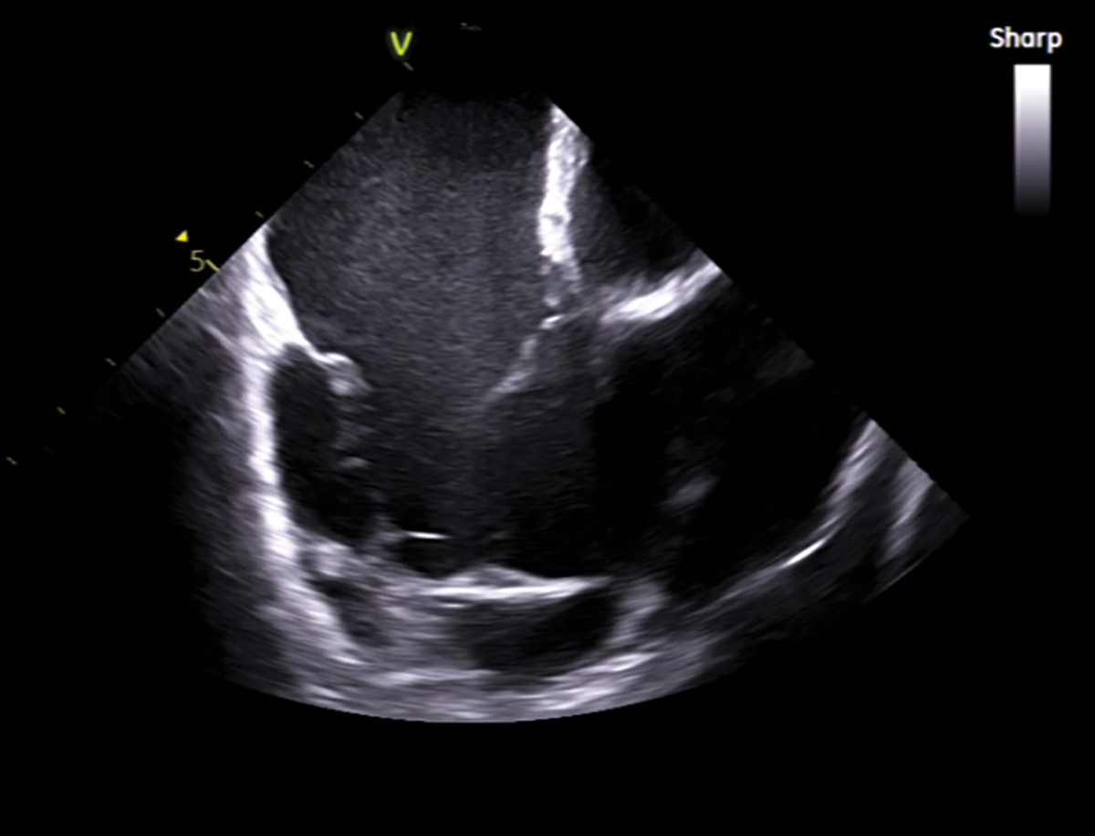
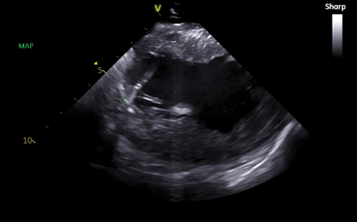
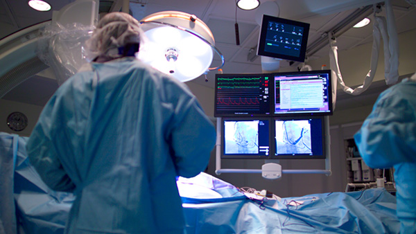
Our workflow includes general anesthesia for all AFib ablations. Importantly, we stopped using lead for our procedures. We rely heavily on the ultrasound images and 3D maps. Ultrasound is used for femoral venous access, significantly decreasing the risk of vascular complications. The ICE catheter is advanced under ultrasound guidance into the right atrium (RA), capturing the "home view" (Figure 1).
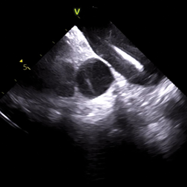
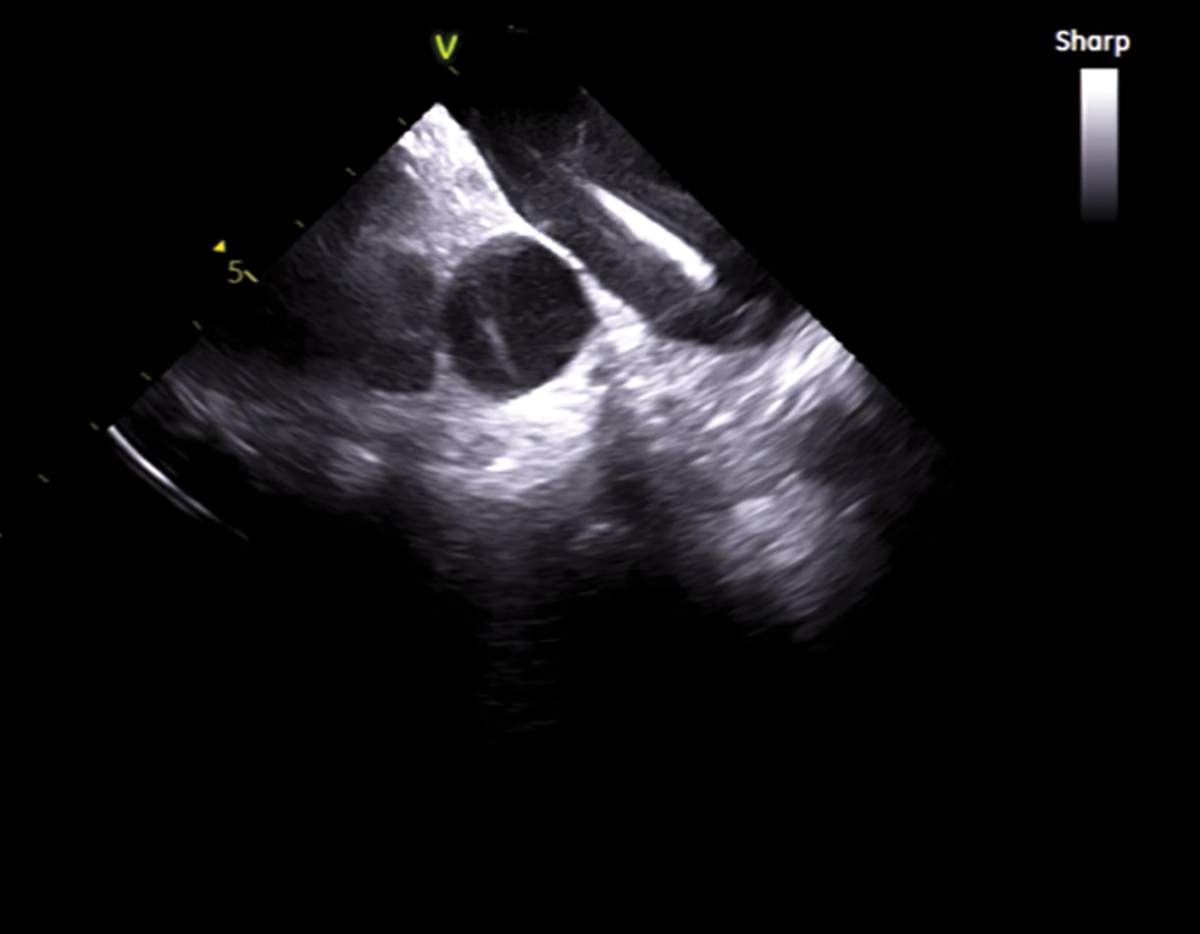
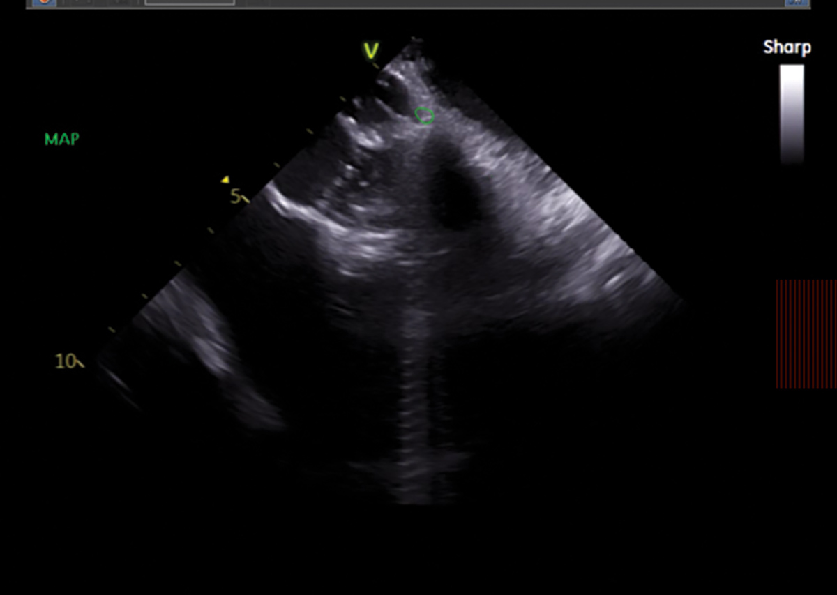
From the home view (ICE catheter in the RA), we perform a baseline assessment of the cardiac chambers, pericardium, valves and intra-atrial septum. A guidewire and steerable sheath are then advanced to the superior vena cava (SVC) (Figure 2).
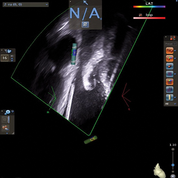
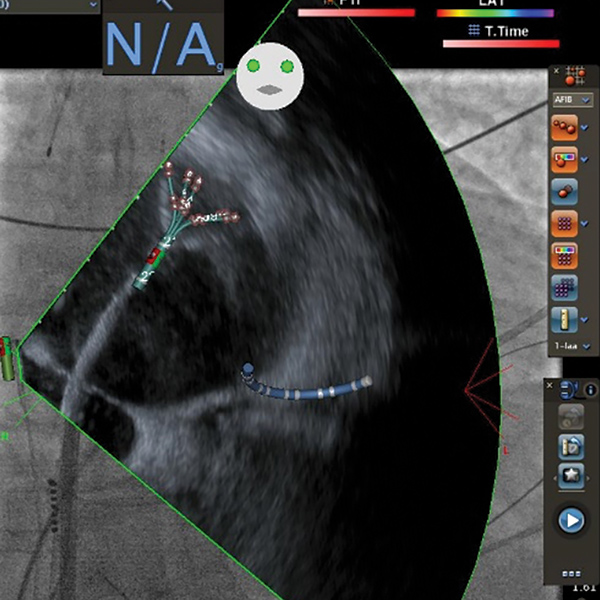
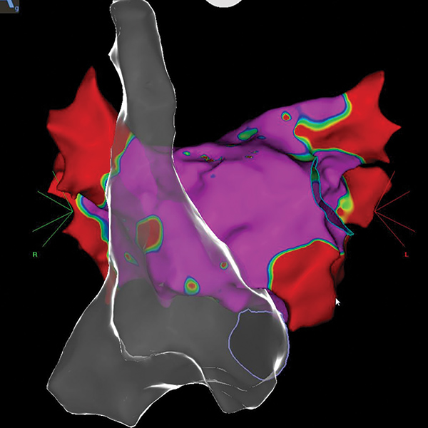
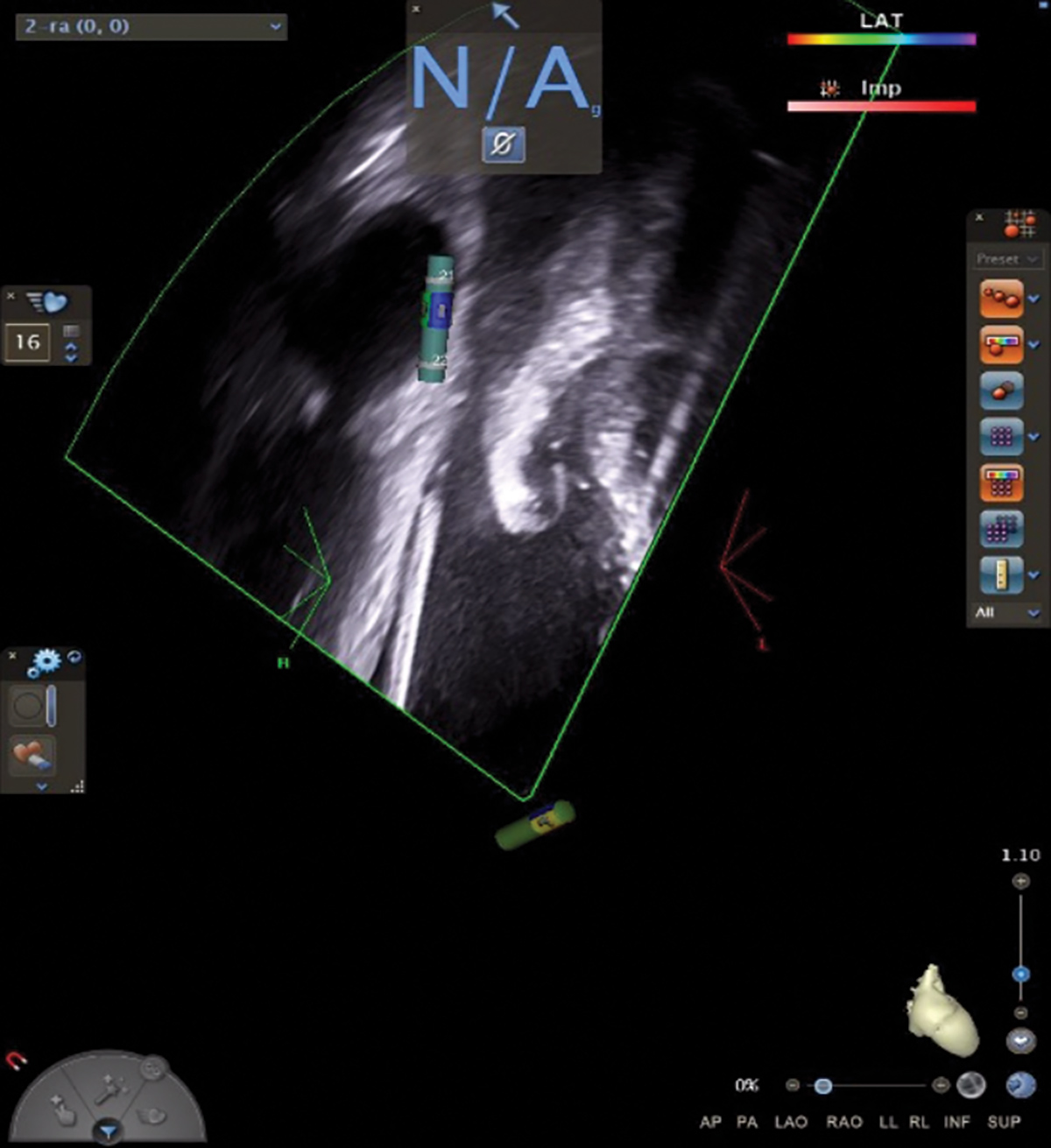
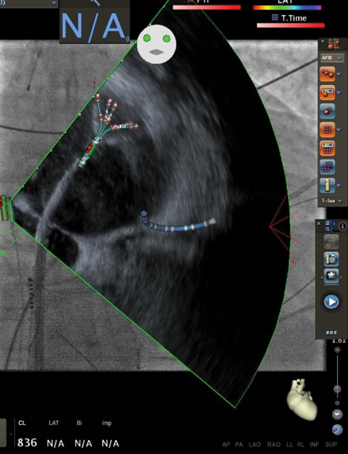
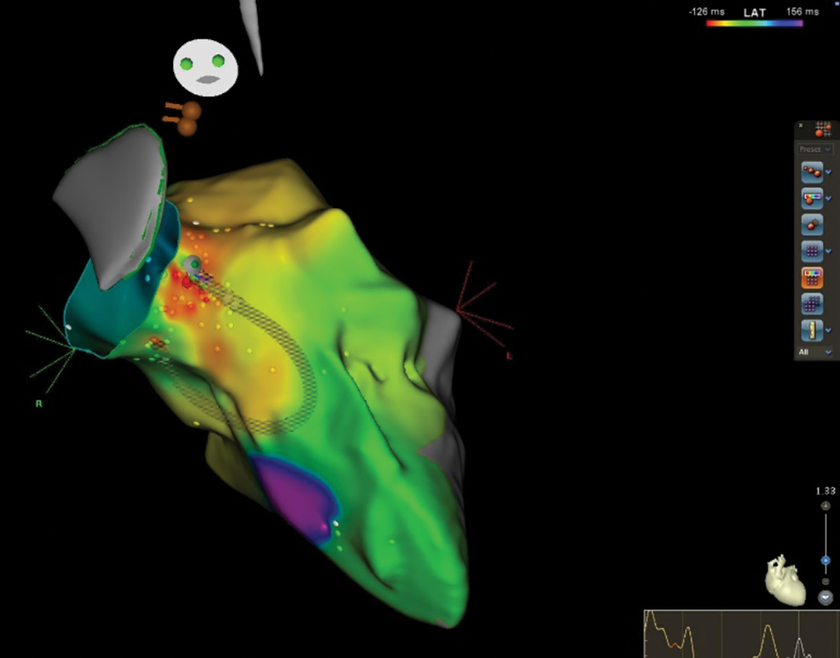
This is followed by a steerable multielectrode mapping catheter (Pentaray; Biosense Webster) that is advanced into the SVC to create the RA geometry and delineate the coronary sinus (CS) on the 3D map (Figures 3, 4).
Subsequently, a steerable decapolar catheter is inserted into the CS under ICE guidance and using the pre-formed RA geometry as a map to guide CS access and aid with catheter placement.
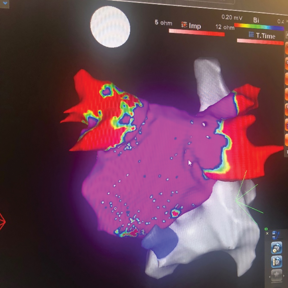
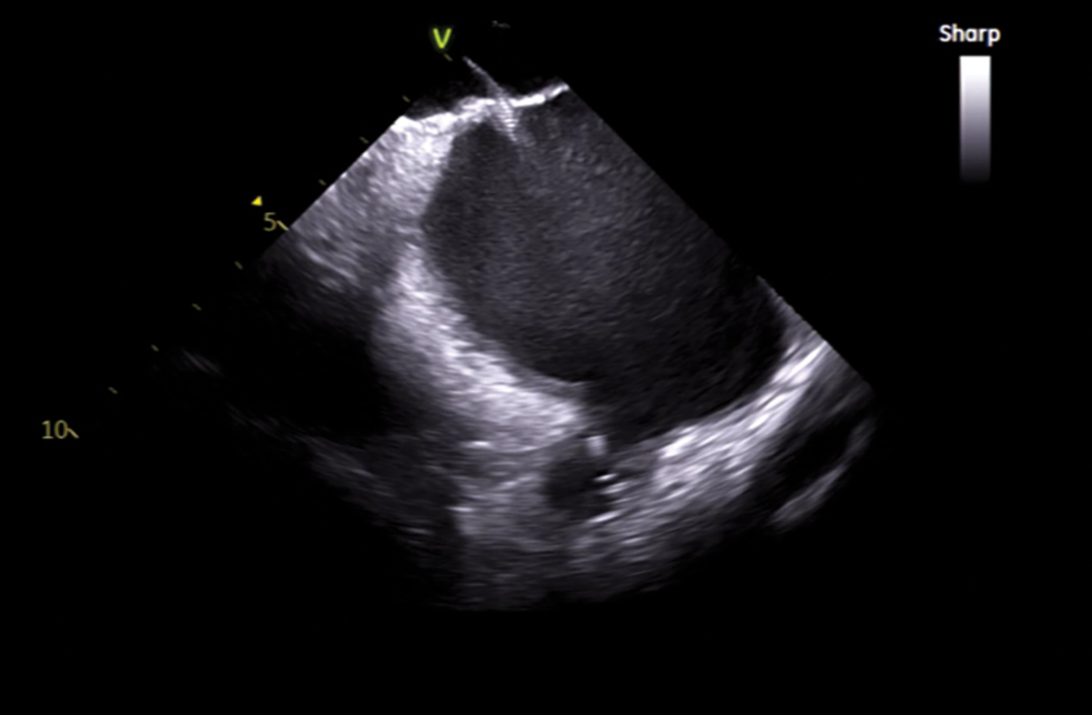
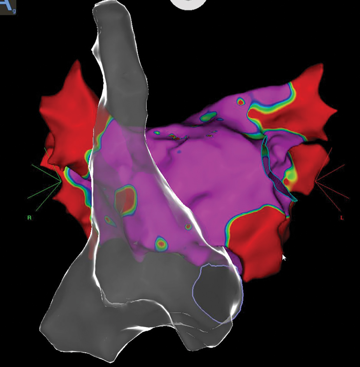
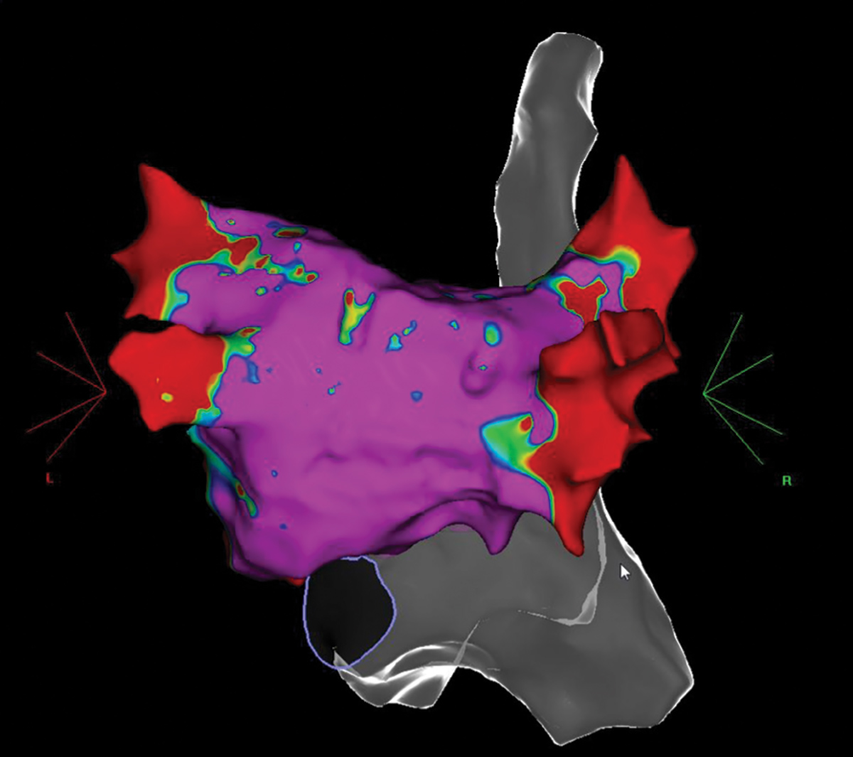
Transseptal puncture is performed under direct ICE guidance (Figure 5). Then the multielectrode mapping catheter is advanced into the left atrium (LA) to create LA geometry (Figure 6), after which it is exchanged for the ablation catheter and ablation is performed.10
Adequate catheter-tissue contact is ensured using a combination of ICE imaging, electrogram characteristics and contact-force sensing. Ablation is performed +/- linear lesions for pulmonary vein isolation, depending on each case and indications.
Using this approach, we've performed zero fluoroscopy AFib ablations with similar safety and efficacy as seen with ablations using fluoroscopy. No increase in procedure times or complications has been seen in our experience.
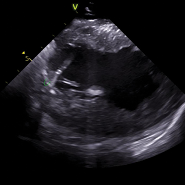
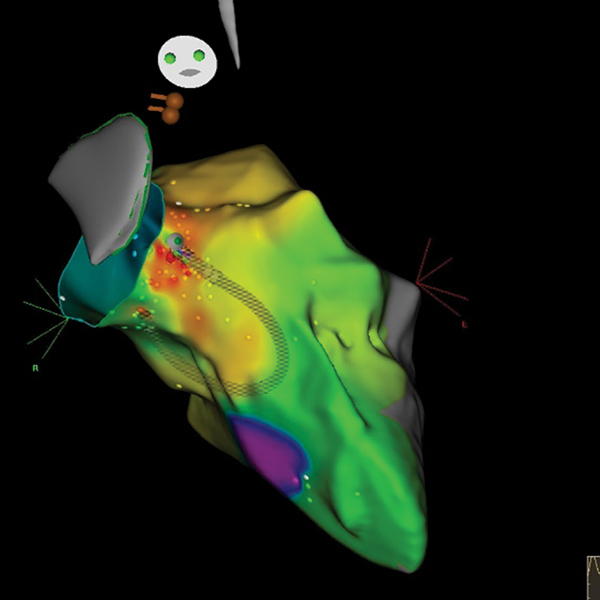
For VAs, we use a similar approach. We rely heavily on the ICE images and image integration of the 3D map using phase-array ICE imaging. This allow us to have a clear and precise visualization of the intracardiac structures, especially papillary muscles, outflow tract, coronary sinuses and surrounding structures (Figures 7, 8).
The approach, retrograde or transseptal, is determined by patient characteristics and risks. For example, in a patient with severe iliofemoral vascular disease and tortuosity, we prefer the transseptal approach. Depending on the location of the target, we select a transseptal vs. retrograde approach.
Recently we reported a case series of 43 patients (30 premature ventricular contractions and 13 VTs) who successfully underwent zero fluoroscopy VT ablation, without compromise to procedure safety or efficacy.11
In summary, the field of EP is advancing at a very fast pace with very promising technology that is making these procedures safer, less time consuming and now with near-zero to non-radiation exposure.
This is definitively a great time to be a cardiologist especially an electrophysiologist.
Fluoroless Procedures for AFib
Click the images below for a larger, more detailed view.
Fluoroless Procedures for VAs
Click the images below for a larger, more detailed view.
References
- Einstein AJ, Henzlova MJ, Rajagopalan S. JAMA 2007;298:317‐23.
- Venneri L, Rossi F, Botto N, et al. Am Heart J 2009;157:118‐24.
- Sarkozy A, De Potter T, Heidbuchel H, et al. Europace 2017;19:1909-22.
- Fernández‐Gómez JM, Moriña‐Vázquez P, Morales ER, et al. J Cardiovasc Electrophysiol 2014;25:638‐44.
- Ferguson JD, Helms A, Mangrum JM, et al. Circ Arrhythm Electrophysiol 2009;2:611‐9.
- Razminia M, Willoughby MC, Demo H, et al. Pacing Clin Electrophysiol 2017;40:425-33.
- Sommer P, Bertagnolli L, Kircher S, et al. Europace 2018;20:1952-8.
- Reddy VY, Morales G, Ahmed H, et al. Heart Rhythm 2010;7:1644‐53.
- Birnie D, Healey JS, Krahn AD, et al. J Cardiovasc Electrophysiol 2011;22:957‐60.
- Sadek MM, Ramirez FD, Nery PB, et al. J Cardiovasc Electrophysiol 2019;30:78-88.
- Johnson A, Mejia-Lopez E, Bilchick K, et al. Catheter ablation of ventricular arrhythmias without fluoroscopy using intracardiac echocardiography and electroanatomic mapping. Presented May 2019 in San Francisco at Heart Rhythm Society Scientific Session.
Clinical Topics: Arrhythmias and Clinical EP, Noninvasive Imaging, Implantable Devices, EP Basic Science, SCD/Ventricular Arrhythmias, Atrial Fibrillation/Supraventricular Arrhythmias, Echocardiography/Ultrasound, Nuclear Imaging
Keywords: ACC Publications, Cardiology Magazine, Pulmonary Veins, Ventricular Premature Complexes, Atrial Fibrillation, Coronary Sinus, Vena Cava, Superior, Atrial Septum, Cardiac Catheters, Papillary Muscles, Fellowships and Scholarships, Learning Curve, Atrial Flutter, Tachycardia, Supraventricular, Catheter Ablation, Heart Atria, Fluoroscopy, Pericardium, Vascular Diseases, Neoplasms, Anesthesia, General, Echocardiography, Electrophysiology
< Back to Listings


