Cryoballoon Pulmonary Vein Isolation
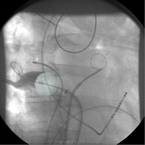
Complete isolation of the pulmonary veins (PV) is considered the cornerstone of catheter intervention to treat atrial fibrillation (AF). PV isolation using a cryoballoon has evolved into a relatively simple alternative for point-by-point radiofrequency current (RFC) ablation because this technology theoretically allows for PV isolation with a single application. The technique has a steep learning curve and can be performed with fluoroscopic guidance without a 3-dimensional mapping system or additional imaging techniques.(1) To achieve continuous lesions, an occlusive balloon position at target PV sites as confirmed by PV angiography is necessary to limit convective heating by leaking blood flow (Figure 1). This may be challenging especially at the right inferior PV due to its proximity to the transseptal puncture site. However, using special catheter maneuvers based on the individual anatomy, complete PV isolation is possible in the great majority of patients (97%) with a single balloon and without additional "touch-up" lesions by a focal cryocatheter.(1) If PV occlusion cannot be achieved, the "pull-down" technique may be used, with freezing initiated at the superior PV circumference, accepting a small inferior leakage. When steady-state balloon temperature is achieved both the sheath and the frozen balloon are pulled down to close the inferior leakage. Successful ablations generally reach a minimal balloon temperature of -40°C or less.(2)
There are currently two available diameters of the Arctic Front™ cryoballoon (Medtronic Cryocath LP), 23 and 28 mm. In our laboratory, we only use the big (28 mm) balloon. The major rationale behind the big cryoballoon strategy is procedural safety by using an intentionally oversized balloon deploying lesions as proximal as possible at the PV antrum. In addition to energy delivery to PV tissue, a deep position inside the vein results in the combined effect of close proximity to adjacent structures and deeper freezing temperatures due to less convective heating of the balloon by atrial blood flow. Accordingly, right-sided phrenic nerve palsy (PNP), the most frequent complication associated with cryoballoon PV isolation, has been reported in a significantly higher rate using the 23mm (12.4%) when compared to the 28mm balloon (3.5%).(3) Although transient in the great majority of patients, PNP may occur due to the close anatomical relationship of the right phrenic nerve to the septal (especially right superior) PVs. In addition to avoiding distal balloon positions, diaphragm movement has to be monitored continuously when freezing at the septal PVs. This is usually performed by high-output pacing of the right phrenic nerve proximal to its intersection with the right superior PV from the superior caval vein (Figure 1, asterisk), and manual palpation of the resultant diaphragm contractions. If weakening of the contractions occurs, cryoablation is immediately stopped. Diaphragmatic electromyography is investigated as a more sensitive tool for the early detection of PNP.(4)Balloon size also plays a role in possible collateral damage to the esophagus. In total, 116 patients have been included in 3 studies with systematic endoscopic screening for esophageal lesions after cryoballoon PV isolation (3). Esophageal ulcerations (6/35, 17%) were only reported in the study using both balloon sizes (and a focal cryocatheter if needed). In the remaining 81 patients from 2 studies employing a single 28mm balloon strategy, no esophageal lesion was found.(3,5) This may be explained by a deviation of the esophageal course from the midline in the great majority of patients, resulting in close proximity of the esophagus to the posteriorly directed inferior PVs. As of today, no atrio-esophageal fistula has been described following cryoballoon ablation, reported esophageal ulcerations healed without clinical sequelae.
A serious complication of PVI using RFC ablation is PV stenosis. Based on the definition of >75% reduction in cross-sectional area from baseline in non-invasive imaging studies, significant PV stenosis was for the first time reported in 7/228 (3.1%) patients following cryoballoon PVI in the North American Arctic Front STOP-AF trial (presented at the Annual Scientific Session in Atlanta, March 2010). Pending publication of the full results, it is unknown whether these cases were associated with a specific balloon size. Energy delivery inside the PV by an under-sized balloon, or "venoplasty" by inflation of an over-sized balloon within the PV are possible explanations. However, combined data from available studies indicate a low incidence (2/1163; 0.17%) of significant PV stenosis resulting in symptoms or requiring intervention.(3)
In patients with paroxysmal AF who underwent cryoballoon PV isolation, one-year freedom from recurrent AF has been reported in the range of 49-77% of patients, depending on whether or not a 3-month blanking period was employed. In persistent AF, one-year freedom from recurrent AF has been reported in 42-48% of patients.(3) Since patients with long-lasting persistent AF may need ablation strategies in addition to PVI to achieve higher success rates, these patients are not treated with cryoballoon ablation in our laboratory. As of today, only data from small non-randomized studies comparing the clinical efficacy of cryoballoon- to RFC-based PVI are available, which showed no difference between the techniques.(3) Sufficiently powered, randomized studies are in preparation.
In conclusion, our approach to cryoballoon PVI using only the single big (28 mm) balloon aims primarily for maximal patient safety. It is a straightforward "single-device" strategy. Large-scale, randomized trials will provide the answer to the important question relating to cryoballoon PVI – whether it constitutes a safer alternative to RFC-based PVI with comparable efficacy.
References
- Kuck K-H, Fürnkranz A. Cryoballoon ablation of atrial fibrillation. J. Cardiovasc. Electrophysiol. 2010;21(12):1427-1431.
- Fürnkranz A, Köster I, Chun KRJ, Metzner A, Mathew S, Konstantinidou M, Ouyang F, Kuck KH. Cryoballoon temperature predicts acute pulmonary vein isolation. Heart Rhythm. 2011, doi:10.1016/j.hrthm.2011.01.044.
- Andrade JG, Khairy P, Guerra PG, Deyell MW, Rivard L, Macle L, Thibault B, Talajic M, Roy D, Dubuc M. Efficacy and Safety of Cryoballoon Ablation for Atrial Fibrillation – A Systematic Review of Published Studies. Heart Rhythm. 2011, doi: 10.1016/j.hrthm.2011.03.050.
- Franceschi F, Dubuc M, Guerra PG, Delisle S, Romeo P, Landry E, Koutbi L, Rivard L, Macle L, Thibault B. Diaphragmatic electromyography during cryoballoon ablation: a novel concept in the prevention of phrenic nerve palsy. Heart Rhythm. 2011, doi:10.1016/j.hrthm.2011.01.031.
- Fürnkranz A, Chun KRJ, Metzner A, Nuyens D, Schmidt B, Burchard A, Tilz R, Ouyang F, Kuck KH. Esophageal endoscopy results after pulmonary vein isolation using the single big cryoballoon technique. J. Cardiovasc. Electrophysiol. 2010;21(8):869-874.
Clinical Topics: Arrhythmias and Clinical EP, Invasive Cardiovascular Angiography and Intervention, Atrial Fibrillation/Supraventricular Arrhythmias, Interventions and Imaging, Angiography, Nuclear Imaging
Keywords: Angiography, Atrial Fibrillation, Electromyography, Esophageal Fistula, Freezing, Heating, Palpation, Patient Safety, Phrenic Nerve, Pulmonary Veins, Punctures, Temperature
< Back to Listings


