Extra Tissue is the Issue
Thank you for visiting ACC.org. Please note that this item was published more than 5 years ago and therefore its content may be outdated. For more current information on this topic, we encourage you to visit our Congenital Heart Disease and Pediatric Cardiology Collection page.
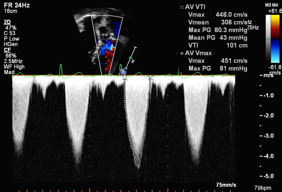
An asymptomatic 43-year-old Ukrainian female presented to the outpatient office for a second opinion. After the birth of her second child in 2005, a murmur was auscultated. A transthoracic parasternal long axis echocardiogram image is shown in Figure 1. Annual echocardiograms revealed a gradual increase in the gradient across the pathology of concern. In March 2015, ten years since the initial presentation, select apical outflow tract images from a follow-up echocardiogram are shown in Figure 2 and Figure 3. A transesopahegeal echocardiogram, a select image of which is shown in Figure 4, confirmed the aforementioned findings.
Figure 1: Transthorcic Parasternal Long axis view (Video)
Figure 2: Transthoracic Apical 5 chamber view (Video)
Figure 3: Pulse Doppler interrogation, Transthoracic Apical 5 chamber view (Image)
Figure 4: Select views from a transesophageal echocardiogram with color Doppler (Video)
What is the next best step for this asymptomatic patient?
Show Answer


