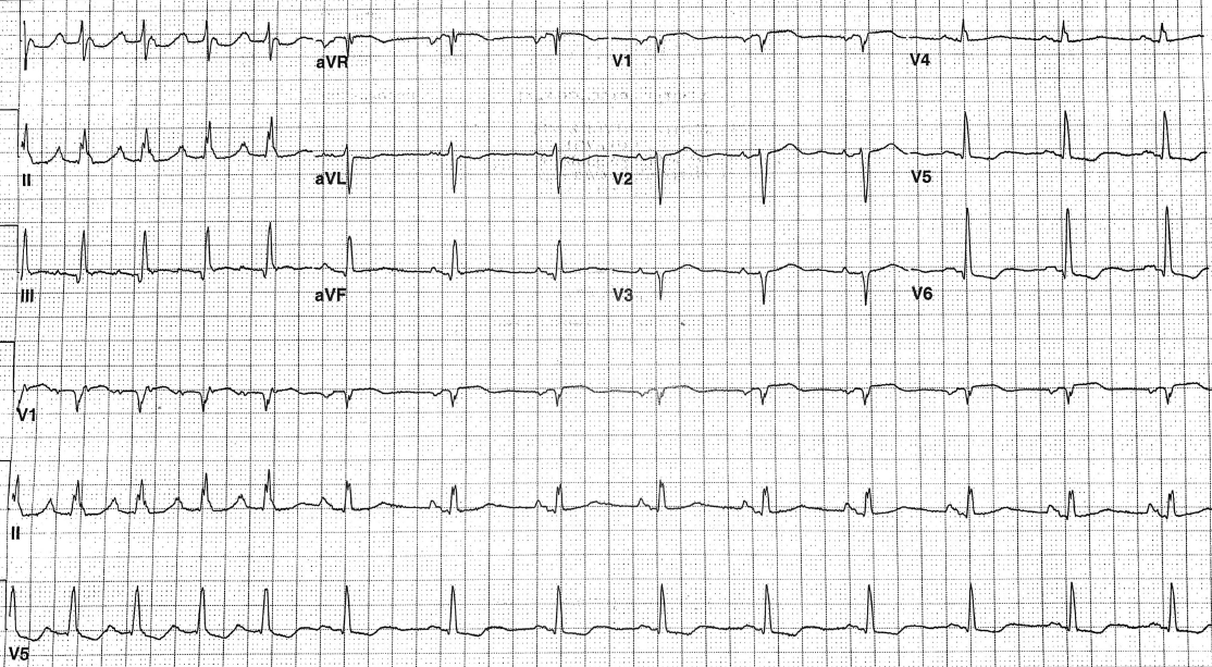
A 59-year-old woman with a history of CABG 10 years ago in the setting of hypertension, hyperlipidemia, and type 2 diabetes mellitus, presents with complaints of palpitations, dyspnea, and weakness. She has had paroxysmal atrial fibrillation for which she is receiving amiodarone. An ECG is performed.
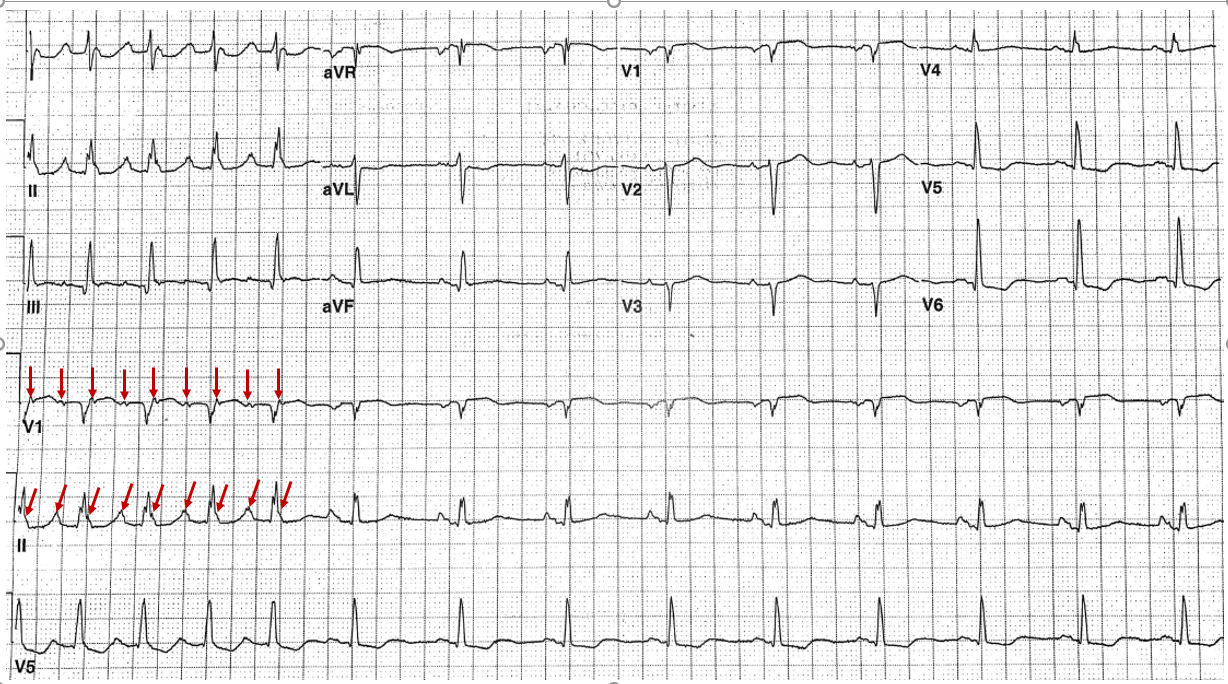
The correct answer is: D. Sinus rhythm with atrial flutter.
After the first five beats, the patient is in sinus rhythm with abnormal P wave secondary to left atrial abnormality.1 The axis is rightward (approximately 105 degrees). The R wave progression is abnormal reflecting the old anterior myocardial infarction. Observation of leads V1 and II (rhythm strips) shows a P wave (flutter wave) in the terminal QRS region as well as a flutter wave proceeding the QRS (red arrows).2
References
- Batra MK, Khan A, Farooq F, Masood T, Karim M. Assessment of electrocardiographic criteria of left atrial enlargement. Asian Cardiovasc Thorac Ann 2018;26:273-6.
- Rodriguez Ziccardi M, Goyal A, Maani CV. Atrial flutter. StatPearls. Treasure Island (FL): StatPearls Publishing 2019.


