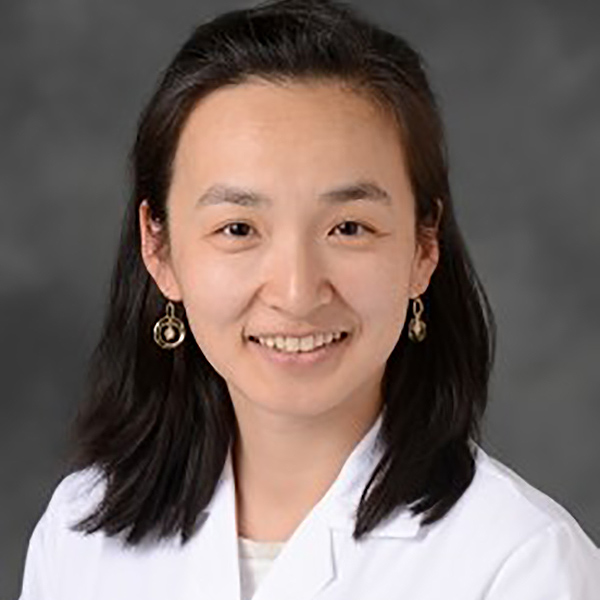
Interview With Dee Dee Wang, MD, FACC

How did you first find your passion for structural cardiac imaging?
Structural heart imaging is a new subspecialty field of cardiology. It is innovative and challenging, and standardized guidelines have yet to be written.
Transcatheter devices are still being conceived by industry and optimal procedure techniques are still being developed. It is an incredible opportunity to be a part of this new field at the ground level.
Tell us about the new Structural Cardiac Imaging Fellowship that you've just started.
The Structural Heart Disease Interventional Imaging Fellowship at Henry Ford Hospital is a one-year non-ACGME accredited advanced fellowship training program available to physicians who have completed training in general cardiology.
The program currently consists of four full-time structural heart interventional cardiologists and two full-time structural heart interventional imaging cardiologists. The program is available to one advanced fellow per year.
The program is designed to train fellows to independently guide non-coronary cardiac interventional procedures including transseptal crossings, percutaneous PFO closure, percutaneous ASD closure, percutaneous VSD closure, percutaneous alcohol septal ablation, percutaneous mitral and aortic balloon valvuloplasty, percutaneous closure of paravalvular leaks, TAVR, TMVR, transcatheter repair for mitral interventions (ie MitraClip), percutaneous left atrial appendage closure, advanced intraprocedural 3D TEE guidance, and advanced peri-procedural Structural CT planning.
Is congenital heart disease imaging a part of your fellowship?
We do not see pediatric congenital heart disease patients. However, we see some adult congenital heart disease patients when they're referred for ASD, VSD, coarctation stenting interventions, PFO closures, pulmonic valve in valve planning, bicuspid aortic valves, or transplant of a patient with a complicated congenital heart anatomy.
You mentioned about 3D printing, could you tell us briefly about it?
The 3D print helps our team in planning high risk transcatheter and surgical procedures. For our fellows, we use 3D printing to teach them anatomy pre-procedurally.
This helps redefine our 2D and 3D TEE imaging for intraprocedural guidance, as well as helps our young cardiologists understand the spatial anatomy present in our intraprocedural images. It is a phenomenal opportunity to teach fellows how to image differently.
What imaging modalities are necessary to master as a structural heart interventional imager?
3D Interventional TEE guidance, structural CT segmentation and basic fluoroscopy are required for structural heart omaging. Structural heart disease imagers must be present at the majority of heart team multidisciplinary conferences reviewing key images with team members to facilitate and lead discussions on patient diagnosis and procedural device anatomical appropriateness.
A lot of places in the country do not have specifically trained people in structural imaging. What is the magnitude of impact we would see by having more specialty trained cardiologists?
JACC: Imaging recently published two Expert Consensus documents led by authors Rebecca T. Hahn, MD, FACC, and Jonathon Leipsic, MD, defining structural imaging core competencies.
The Journal of Structural Heart published our teams' expert opinion recommendations for minimal training requirements for structural heart imagers.
The impact of structural heart disease imaging is already being experienced by the field of cardiology with a call for recognition of these new skillsets and necessary training requirements.
There is a shortage of structural heart imaging cardiologists. As surgical volumes become increasingly absorbed by transcatheter interventions, there will be a greater need for structural imagers.
Would it impact outcomes?
It has been well-documented and studied that there is an early-operator learning curve to procedural competency. There is now even a minimum credentialing and competency requirement in TAVR operators.
The same learning curve not only exists in interventional imaging for structural heart disease, but the degree of learning is even more amplified.
Traditional imagers asked to step in to the role of interventional imaging may have had little recent exposure to or set foot into the sterile field of a cardiac catheterization lab, which is a significant contrast to interventional cardiologists who are asked to transition to become structural heart implanters.
In a recent JACC publication by Adnan Chhatriwalla, MD, FACC, et al., there was clear demonstration that "operator experience was associated with improvements in procedural success, procedure time and procedural complications. The effect of operator experience was greater when considering the goal of achieving 1+ residual mitral regurgitation."
How much is radiation exposure a concern?
Structural imagers receive three times the radiation exposure of an interventionalist in the same procedure. At Henry Ford hospital, we are very careful with our radiation shielding and there is significant administrative support to make that possible. We hope that would be the standard for every imager across the country and we try to encourage that.
Where do you think this field is headed?
If you want to be part of transcatheter therapy innovations, this is a great field to pursue. However, it is not heading anywhere if we do not have people training for it. We need formalized training programs and reimbursement models to allow this field to thrive.
Any advice to people who are considering training in structural heart imaging?
Apply to Henry Ford Hospital. Apply, learn it, love it and enjoy the ride.


