The umbilical vein is seen connecting directly to the right atrium (RA) with unrestricted flow in Videos 1 and 2. Labeled frames from Video 1 are shown in Figure 1 and from Video 2 in Figure 2.
Figure 1
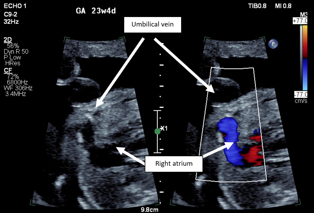 Figure 1: Two-Dimensional and Corresponding Color Doppler Images in an Axial Plane Demonstrating the Umbilical Vein Connecting Directly to the Right Atrium. Courtesy of Crafts LS, Powell AJ.
Note that the umbilical vein color Doppler flow is easily seen even though the Nyquist limit is set relatively high for venous flow at 77 cm/sec; this is a sign that flow may be increased and should prompt further investigation.
Figure 2
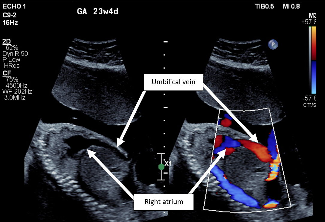 Figure 2: Two-Dimensional and Corresponding Color Doppler Images in a Sagittal Plane Demonstrating Umbilical Vein Flow Through the Liver Without Restriction Before Entry into the Right Atrium. Courtesy of Crafts LS, Powell AJ.
The fetus has qualitatively moderate to severe cardiomegaly with dilatation of all four chambers (Image 1).
In the fetal circulation, oxygen and nutrient-rich blood are delivered to the fetus via the umbilical vein. The ductus venosus acts as a shunt and a restrictor, allowing some umbilical venous blood flow to bypass the liver. Instead of traveling through the portal venous system, 20-30% of umbilical venous blood flows from the umbilical vein to the left portal vein to the ductus venosus to the IVC. This IVC blood then preferentially streams across the foramen ovale to the left heart, thereby delivering oxygen and nutrient-rich blood to the cerebral and coronary circulations.
The ductus venosus regulates flow between the higher-pressure umbilical venous system and the lower-pressure systemic venous system. It is an active structure, such that during periods of hypoxemia and hypovolemia, the ductus venosus dilates to maintain flow and sustain oxygen delivery to the cerebral and coronary circulations at the expense of portal venous flow.1
There are many types of congenital anomalies of the umbilical-portal venous system. The type shown in this case is called an umbilical vein–to–systemic venous shunt. In this category of anomalies, there is likely agenesis of the left portal vein and the ductus venosus. Instead, blood drains from the umbilical vein directly to the systemic venous circulation at sites such as the RA (as in this case), IVC, renal vein, iliac vein, left atrium, or coronary sinus.2 This anatomy creates a direct connection between the umbilical system (higher pressure) and systemic venous system (lower pressure). Without the ductus venosus acting as a resistor, regulating the amount of blood flow bypassing the liver, there can be increased blood flow that bypasses the liver and enters the systemic venous circulation.3 The increased volume load to the heart may cause dilatation of the atria and ventricles (cardiomegaly), atrioventricular valve dysfunction, and ventricular dysfunction. In severe cases, fetal hydrops may occur secondary to high-output cardiac failure, and early delivery may be required. Outcomes of fetuses with an absent ductus venosus and extrahepatic umbilical venous drainage, as in this case, are largely determined by the presence or absence of hydrops.4,5 Other congenital abnormalities have been associated with umbilical-systemic shunts including chromosomal, facial, vertebral, cardiac, genitourinary, and gastrointestinal tract anomalies.5 Up to 25% of infants with absence of the ductus venosus can have abnormalities of portal venous drainage. Therefore, postnatal assessment by hepatology may be warranted.
The fetus in this case had moderate to severe cardiomegaly, normal valvular function, and normal ventricular function. The fetus was monitored closely with serial ultrasounds and fetal echocardiograms. The infant was born at term via uncomplicated spontaneous vaginal delivery. A postnatal echocardiogram had findings of mild mitral regurgitation, otherwise normal valvular function, biventricular dilatation, and normal ventricular function. A follow-up echocardiogram at 6 months of age had findings of normal ventricular size and function.
References
- Kiserud, T, Kessler J. The ductus venosus. In: Maulik D, Lees CC, eds. Doppler Ultrasound in Obstetrics and Gynecology. Cham, Switzerland: Springer; 2023.
- Qin Y, Wen H, Liang M, et al. A new classification of congenital abnormalities of UPVS: sonographic appearances, screening strategy and clinical significance. Insights Imaging 2021;12:125.
- Gorincour G, Droullé, P, Guibaud L. Prenatal diagnosis of umbilicoportosystemic shunts: report of 11 cases and review of the literature. AJR Am J Roentgenol 2005;184:163-8.
- Kiserud T. Hemodynamics of the ductus venosus. Eur J Obst Gynecol Reprod Biol 1999;84:139-47.
- Berg C, Kamil D, Geipel A, et al. Absence of ductus venosus-importance of umbilical venous drainage site. Ultrasound in Obstet Gynecol 2006;28:275-81.


